Archaeologists found evidence of trepanation on medieval woman’s skull
[ad_1]

I. Micarelli et al., 2023
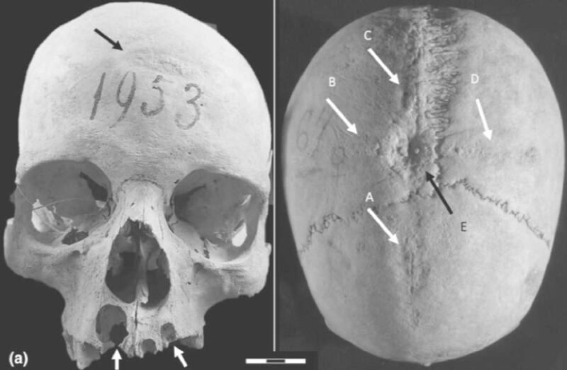
Scientists analyzed the skull of a medieval woman who once lived in central Italy and found evidence that she experienced at least two brain surgeries consistent with the practice of trepanation, according to a recent paper published in the International Journal of Osteoarchaeology. It’s one of the few archaeological pieces of evidence of trepanation being performed on early medieval women yet found, although why the woman in question was subjected to such a risky invasive surgical procedure remains speculative.
Archaeologists have found evidence of various examples of primitive surgery dating back several thousand years. For instance, last year, archaeologists excavated a 5,300-year-old skull of an elderly woman (about 65 years old) from a Spanish tomb. They determined that seven cut marks near the left ear canal were strong evidence of a primitive surgical procedure to treat a middle ear infection. The team also identified a flint blade that may have been used as a cauterizing tool. By the 17th century, this was a fairly common procedure to treat acute ear infections, and skulls showing evidence of a mastoidectomy have been found in Croatia (11th century), Italy (18th and 19th centuries), and Copenhagen (19th or early 20th century).
Cranial trepanation—the drilling of a hole in the head—is perhaps the oldest known example of skull surgery and one that is still practiced today, albeit rarely. It typically involves drilling or scraping a hole into the skull to expose the dura mater, the outermost of three layers of connective tissue, called meninges, that surround and protect the brain and spinal cord. Accidentally piercing that layer could result in infection or damage to the underlying blood vessels. The practice dates back 7,000 to 10,000 years, as evidenced by cave paintings and human remains. During the Middle Ages, trepanation was performed to treat such ailments as seizures and skull fractures.
The skull examined for this latest paper was excavated in the late 19th century from the Longobard cemetery of Castel Trosino in central Italy, dated between the 6th and 8th centuries CE, along with hundreds of other burials. But only 19 skulls found at the site were in sufficiently good condition for study. Castel Trosino was strategically important to the region in the wake of the collapse of the Roman Empire. According to the authors, the Byzantine government wanted to stabilize its control of central Italy and ensure peace by encouraging members of prestigious Longobard families to live there. So the burial sites included gold objects and fine jewelry along with the human remains.

I. Micarelli et al., 2023
This particular skull is that of a woman about 50 years old. It was found in a double burial, along with male remains, plus a bronze brooch, comb, and gold filaments that suggested the couple belonged to one of those elite families. The authors initially examined the skull and noted several cranial defects—specifically, a cross-shaped section of porous bone with an oval-shaped depression at the center and a second oval-shaped scraped area.
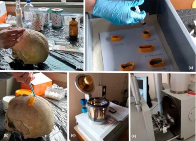
So they took CT scans of the skull and made casts to learn more. They cleaned the bone surface with cotton swabs and let it dry before applying blue silicone to the areas with bone defects with a dispensing gun. Then a second layer of orange silicone was added to cover the blue silicone. Then casts were made of epoxy resin and left to dry for 48 hours. The cast surfaces were then metallicized with golden powder and scanning electron microscopy was used to analyze the samples.
“We found that the woman had survived several surgeries, having undergone long-term surgical therapy, which consisted of a series of successive drillings,” said co-author Ileana Micarelli of the University of Cambridge and a former postdoc at Sapienza University, which coordinated the international study.
[ad_2]
Source link


